Photosynthesis Overview

Overview of Photosynthesis:

Photosynthesis involves the conversion, by various phototrophic organisms, of light energy into organic molecules, with or without the production of oxygen.

Nonoxygenic, anaerobic photosynthesis evolved^ in prokaryotes before oxygenic photosynthesis, and is found in green filamentous, green sulfur, purple sulfur, and purple nonsulfur bacteria. Photosynthesis in green sulfur bacteria such as Chlorobium tepidum takes place in the chlorosome.

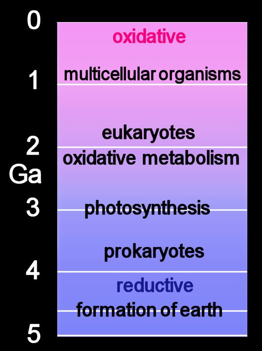
Oxygenic photosynthesis apparently developed several billion years ago in an ancestor of present day Cyanobacteria. Oxygenic photosynthesis occurs in plants, which possess plastids derived from Cyanobacteria, and in photosynthetic oxygenic prokaryotes, which utilise H2O as electron donor (Cyanobacteria, prochlorophytes).

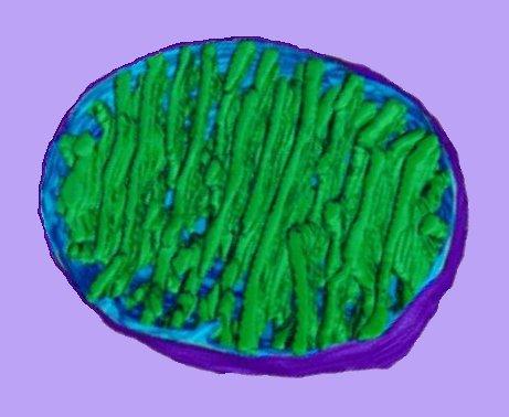
The photosystem machinery of oxygenic photosynthesis is located within the specialized internal thylakoid membrane system. The chloroplast is the site of oxygenic photosynthesis in eukaryotic cells.

The current consensus is that chloroplasts originated from Cyanobacteria that have become endosymbionts. This is an origin analogous to the endosymbiotic (im) origin of mitochondria, which are believed derived from the "purple bacteria" (alpha-proteobacteria).

The thylakoid membrane, with its embedded membrane-bound pigments, is the structural unit of photosynthesis. Both photosynthetic prokaryotes and photosynthetic eukaryotes possess membranes with embedded photosynthetic pigments. Only eukaryotes, which have a nuclear membrane and membrane-bound organelles, have chloroplasts with an encapsulating membrane.

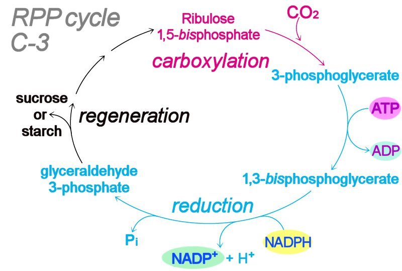
Light-dependent photosynthetic reactions employ the thylakoid membrane-embedded antenna system to harness energy delivered by a photon. The light-dependent, photophosphorylation reactions of photosystems ultimately transduce the energy of light to generate molecules of ATP and NADPH, which act as energy-transfer molecules in the light independent (“dark") reactions of the Calvin cycle. Table ~ comparison photosynthesis & respiration : Table ~ comparison plant bacterial photosynthesis : : Table ~ comparison of C-3, C-4, CAM plants :

The overall reaction of oxygenic photosynthesis is:

6 CO2 + 12 H2O → C6H12O6 + 6 O2 + 6 H2O
or,
CO2 + 2 H2O = CH2O + H2O + O2

Microbial Photosynthesis
Cyanobacteria and prochlorophytes conduct oxygenic photosynthesis. Several photosynthetic bacteria, such as the sulfur bacteria, do not generate oxygen. Nonoxygenic photosynthesis, or cyclic photophosphorylation differs from oxygenic photosynthesis in several ways other than lack of oxygen production.

Hydrogen sulfide (H2S) is utilized by the purple sulfur and green sulfur bacteria:

CO2 + 2H2S = CH2O + H2O + 2So

Nonoxygenic photosynthesis in prokaryotes, utilizing S– or So or H2 as electron donor include green filamentous, green sulfur, purple sulfur, and purple nonsulfur.

Tables  Overview of Photosynthesis  Comparison of C-3, C-4, CAM plants  Comparison of plant and bacterial photosynthesis  Structure of bacteriochlorophylls  Comparison of Photosynthesis and Respiration

Diagram  Z-scheme of noncyclic photophosphorylation :


HOME • • Section PhotosynthesisCalvin cycleC-3C-4CAMChloroplastChlorosomesCyanobacterial cell : cyclic photophosphorylation : • Light-reactions : noncyclic photophosphorylation : • Nonoxygenic photosynthesisOxygenic photosynthesisPhotosynthesis OverviewPhotophosphorylationPlant cellTimeline • Section PigmentsAntenna and Reaction CenterBacteriochlorophyllsCarotenoidsChlorophylls and accessory pigmentsPigments and absorption spectraPhycobilins Section ArticlesSITE MAP • : photosynthesis ~ animations :

Photophosphorylation

Photophosphorylation is the light dependent process by which a proton gradient generates ATP from ADP and Pi.

ADP + Pi →ATP synthase→ ATP

The process of generating a proton gradient resembles that of the electron transport chain of respiration.

Photophosphorylation may be cyclic or noncyclic:

In cyclic photophosphorylation, excited electrons resulting from the absorption of light in Photosystem I are received by the primary electron acceptor and then transferred to the cytb6-f complex, which acts as an electron transport chain. The electrons are returned back to the reaction center of Photosystem I. The excited electrons of cyclic photophosphorylation generate the proton gradient that the ATPase employs to synthesize ATP. No reduction of NADP+ occurs in cyclic photophosphorylation.

In noncyclic photophosphorylation, ATP is generated by the protons gradient created across the thylakoid membranes during the Z-scheme (diagram). The Cytochrome b6-f complex acts as an electron transfer chain. As the electrons release energy during a series of redox reactions, protons are pumped into the thylakoid space. This proton gradient is used for chemiosmotic generation of ATP. The excited electrons are passed on to Photosystem II, where an extra photon of light is harnessed for the reduction of NADP+.

Image :

HOME • • Section PhotosynthesisCalvin cycleC-3C-4CAMChloroplastChlorosomesCyanobacterial cell : cyclic photophosphorylation : • Light-reactions : noncyclic photophosphorylation : • Nonoxygenic photosynthesisOxygenic photosynthesisPhotosynthesis OverviewPhotophosphorylationPlant cellTimeline • Section PigmentsAntenna and Reaction CenterBacteriochlorophyllsCarotenoidsChlorophylls and accessory pigmentsPigments and absorption spectraPhycobilins Section ArticlesSITE MAP

Light-reactions

The photosynthetic reactions that require light occur within the thylakoid membrane. These light-dependent photosynthetic reactions employ the thylakoid membrane-embedded antenna system to harness energy delivered by a photon. The light-dependent, photosystem reactions ultimately transduce the energy of light to generate molecules of ATP and NADPH, which act as energy-transfer molecules in the light-independent, “dark” reactions of the Calvin cycle.

The thylakoid-embedded antenna complex comprises closely packed pigment molecules and the reaction center. Any of the pigment molecules can be excited by a photon of light, and pass their energy along to nearby pigment molecules until the excitation eventually reaches a specialized molecule of chlorophyll called the reaction center, which delivers an excited electron to the electron acceptor molecule in an electron transfer chain. The antenna, including the reaction center, and the electron transport molecules together make up a photosystem.

There are two kinds of photosystems in eukaryotes – PSI and PSII. The reaction center chlorophyll (im) molecule within the antenna of photosystem I responds most strongly to 700 nm light, and is therefore termed P700. The reaction center within the antenna of photosystem II responds most to 680 nm light, and is accordingly called P680. Photosystem I evolved very early, and it is found in nonoxygenic phototrophs; photosystem II evolved later. Because the PSII photosystem is most sensitive to shorter wavelength 680 nm light, it absorbs slightly more energy than the P700-PSI system.

The electron transport system of each photosystem is embedded within the thylakoid membrane and functions in the production of ATP. The system comprises membrane-bound electron carriers that pass electrons from one molecule to the next.

Water is split to generate an electron, hydrogen ions, and oxygen:
2H2O → 4e- + 4H+ + O2

Mechanism of Photophosphorylation
By receiving the energized electron (reduction), the first carrier of the P680 (PSII) electron transport system gains energy. It utilizes some of the energy to pump H+ into the thylakoid lumen, then passes the less energetic electron to the second of four carrier molecules. Each successive carrier in the electron transport chain utilizes some of the energy of the received electron to pump H+ from the stroma into the thylakoid lumen, and then passes the further depleted electron along to the next carrier. Thus, the electron transport system functions to generate a concentration gradient of H+ inside the thylakoid. The chemical potential energy of the H+ concentration gradient is employed to synthesize ATP.

In the process called photophosphorylation, ATP synthase produces ATP from ADP and Pi when hydrogen ions pass out of the thylakoid. The electron, with its energy almost spent, is passed to the P700 antenna of the PSI photosystem and a second electron transport chain. The P680-PSII system has thus generated ATP, a H+ concentration gradient, and energy (Z-scheme (image) of noncyclic photophosphorylation).

The antenna of the evolutionarily older PSI system absorbs a photon of light, and, like the PSII antenna, passes the energy along to the PSI reaction center. Like the PSII system, the P700 reaction center passes the energized electron to an electron acceptor. However, unlike noncyclic photophosphorylation, the electron is retained in the PSI system (cyclic photophosphorylation). So, unlike the PSII system, the electron acceptor of the PSI system utilizes the delivered energy to reduce only molecules of NADP+ to NADPH.

The “light” reactions of noncyclic photophosphorylation produce both ATP and NADPH, which act as energy-transfer molecules in the light-independent (“dark”) reactions of the Calvin cycle. The “dark”, or light-independent Calvin anabolic reactions occur in the stroma of the chloroplast in either light or dark conditions. The light-independent reactions function to reduce CO2 to glucose:

6CO2 + 6H2O → Energy + C6H12O6 + 6O2.

The manganese-calcium oxide cluster, also referred to as the "Oxygen Evolving Complex", OEC, or photosynthetic water oxidase. The OEC is located on the oxidizing side of Photosystem II (PSII).

HOME • Section Photosynthesis • Items alphabetic • Calvin cycleChloroplastChlorosomesCyclic photophosphorylationLight-reactionsPhotosynthesisNoncyclic photophosphorylationNonoxygenic photosynthesisOxygenic photosynthesisPhotophosphorylation • • Section PigmentsAntenna and Reaction CenterBacteriochlorophyllsCarotenoidsChlorophylls and accessory pigmentsPigments and absorption spectraPhycobilins • Section ArticlesSITE MAP

External link Light-reactions :

Labels: , , , , , , , , ,

Nonoxygenic photosynthesis

Several photosynthetic bacteria, such as the sulfur bacteria, do not generate oxygen. Nonoxygenic, or aerobic photosynthesis differs from oxygenic photosynthesis in other ways than lack of oxygen production:

1) Pigments called bacteriochlorophylls (bacteriochlorophyll-a and carotenoids in purple bacteria) absorb light of longer wavelengths than the chlorophylls and accessory pigments of oxygenic Cyanobacteria or photosynthetic eukaryotes.
2) Reduced compounds such as hydrogen sulfide or organic molecules provide the necessary electrons for the reduction of carbon dioxide. (The oxygenic phototrophs employ reduced oxygen – water – as electron donor for reduction of CO2.)
3) The purple bacteria utilize only one photosystem, PSI, while oxygenic phototrophs utilize two photosystems (PSI and PSII).
4) Photophosphorylation is cyclic in nonoxygenic photosynthesis.

The photosynthetic machinery of nonoxygenic photosynthetic purple bacteria is often located in intracytoplasmic membranes. It is not yet known whether or not these membranes are similar to the thylakoid membrane of oxygenic phototrophs, or whether these intracytoplasmic membranes of nonoxygenic phototrophs are merely an extension of the plasma membrane.

HOME • • Section PhotosynthesisCalvin cycleC-3C-4CAMChloroplastChlorosomesCyanobacterial cell : cyclic photophosphorylation : • Light-reactions : noncyclic photophosphorylation : • Nonoxygenic photosynthesisOxygenic photosynthesisPhotosynthesis OverviewPhotophosphorylationPlant cellTimeline • Section PigmentsAntenna and Reaction CenterBacteriochlorophyllsCarotenoidsChlorophylls and accessory pigmentsPigments and absorption spectraPhycobilins Section ArticlesSITE MAP

External link Nonoxygenic photosynthesis

Oxygenic photosynthesis

Oxygenic photosynthesis utilizes light energy to generate ‘excited’ electrons from water:
2H2O → 4e- + 4H+ + O2

A photon is absorbed by Photosystem II and the two resultant excited electrons are passed to Photosystem I, which employs a second photon to further boost their energy for the overall reaction: NADP+ + H+ + 2e- → NADPH.

Thus, the excited electron is subsequently passed to an electron acceptor rather than cycling as in nonoxygenic photophosphorylation. Photophosphorylation is the process of creating ATP using a proton gradient created by the energy gathered from sunlight

Oxygenic photosynthesis by photosystem II (P680-PSII) incorporates photobiochemical capacity specific to cyanobacteria and chloroplasts. Protons (H+) are pumped into the thylakoid lumen, generating a concentration gradient that induces thylakoid ATP synthase to phosphorylate ADP to the energy-storage moiety ATP.

ADP + Pi →ATP synthase ATP

Utilization of proton movement to join ADP and Pi is termed chemiosmosis. This is accomplished by enzymes called ATP synthases or ATPases.

Diagram • Z-scheme of noncyclic photophosphorylation :

Oxygenic photosynthesis apparently developed several billion years ago in an ancestor of present cyanobacteria. The oxygenic photosynthetic machinery is located within the specialized internal thylakoid membrane system. The ability to construct and modify this thylakoid membrane system appears to be an important feature of oxygenic photosynthesis.

HOME • Section Photosynthesis • Items alphabetic • Calvin cycleChloroplastChlorosomesCyclic photophosphorylationLight-reactionsPhotosynthesisNoncyclic photophosphorylationNonoxygenic photosynthesisOxygenic photosynthesisPhotophosphorylation • • Section PigmentsAntenna and Reaction CenterBacteriochlorophyllsCarotenoidsChlorophylls and accessory pigmentsPigments and absorption spectraPhycobilins • Section ArticlesSITE MAP

External link Oxygenic photosynthesis

C-3

Left (click to enlarge):
The C-3, Calvin, photosynthetic carbon reduction (PCR), reductive pentose phosphate cycle (RPP), carbon fixation.

This reaction takes place in the leaves of most plants. During C-3, carbon is fixed, reduced, and utilized via the formation of intermediate 3-carbon sugar phosphates in a cyclic sequence. One complete RPP cycle incorporates three molecules of carbon dioxide and produces one molecule of the three-carbon compound glyceraldehyde 3-phosphate.

The C-3 pathway proceeds in three stages:
1. CO2 fixation (carboxylation) by Rubisco,
2. Carbon reduction to (CH2O)
12 PGA + 12 ATP -> 12 bisPGA + 12 NADPH + 12 H+ -> 12 GAP + 12 NADP+ + 12 Pi, and
3. Regeneration of the CO2 acceptor moleucle (ribulose 1,5-bisphosphate).

Some of the glyceraldehyde 3-phosphate generated in the reductive stage undergoes gluconeogenesis to form glucose. In plants, glucose is converted to sucrose or starch for later use.

The C-3 reactions are sometimes called the "dark reactions" of photosynthesis because photon energy is not used directly – the reactions are light-independent. However, ATP and NADPH generated by light-dependent photophosphorylation reactions are required.

Table ~ comparison of C-3, C-4, CAM plants :

The reaction cycle employs combinations of different length sugar-phosphates and eventually regenerates RuBP in addition to sugar-phosphate for sucrose/starch synthesis.
3-GAP (3 C) → DHAP (3 C)
DHAP (3C) + GAP (3 C) → fructose-1,6-bisphosphate (6C)
fructose-1,6-bisP (6C) + H2O → fructose-6-P (6C) + Pi
fructose-6-P (6 C) + GAP (3 C) → Xylulose-5-phosphate (5 C) + erythrose-4-phosphate (4 C)
erythrose 4-P (4 C) + DHAP (3 C) → sedoheptulose 1,7-bisphosphate (7 C)
sedoheptulose 1,7-bisP + H2O → sedoheptulose 7-P + Pi
sedoheptulose 7-P (7 C) + GAP (3 C) → xylulose 5-phosphate (5 C) + ribose 5-phosphate (5 C)
xylulose 5-P or ribose 5-P → ribulose 5-P
ribulose 5-P + ATP → RuBP (more)

HOME • • Section PhotosynthesisCalvin cycleC-3C-4CAMChloroplastChlorosomesCyanobacterial cell : cyclic photophosphorylation : • Light-reactions : noncyclic photophosphorylation : • Nonoxygenic photosynthesisOxygenic photosynthesisPhotosynthesis OverviewPhotophosphorylationPlant cellTimeline • Section PigmentsAntenna and Reaction CenterBacteriochlorophyllsCarotenoidsChlorophylls and accessory pigmentsPigments and absorption spectraPhycobilins Section ArticlesSITE MAP

Calvin cycle

The Calvin cycle is a light independent ("dark-reaction") process of carbon fixation that takes place in the chloroplasts of all plants, whether C3, C4, or CAM plants. The Calvin cycle utilizes light energy stored as ATP and NADPH to convert CO2 and H2O to organic compounds.

6 CO2 + 12 NADPH + 12 H+ + 18 ATP → C6H12O6 + 6 H2O + 12 NADP+ + 18 ADP + 18 Pi

Enzymes of the Calvin cycle are functionally equivalent to many enzymes involved in other metabolic pathways such as glycolysis and gluconeogenesis, but they are located in the chloroplast stroma rather than in the cytoplasm, thus functionally separating the reactions.

The enzymes are activated by light (hence light independent rather than "dark reaction") and by products of the light-dependent photosynthetic reactions. These regulatory mechanisms prevent the Calvin cycle from operating in reverse to respiration, thus preventing a continuous cycle of CO2 reduction to carbohydrates from occurring simultaneously with carbohydrate oxidation to CO2 (respiration). This regulation prevents the waste of energy (as ATP) in simultaneous reverse reactions that would have no net productivity.

Table ~ comparison photosynthesis & respiration : Table ~ comparison plant & bacterial photosynthesis : Table ~ comparison of C-3, C-4, CAM plants :

HOME • • Section PhotosynthesisCalvin cycleC-3C-4CAMChloroplastChlorosomesCyanobacterial cell : cyclic photophosphorylation : • Light-reactions : noncyclic photophosphorylation : • Nonoxygenic photosynthesisOxygenic photosynthesisPhotosynthesis OverviewPhotophosphorylationPlant cellTimeline • Section PigmentsAntenna and Reaction CenterBacteriochlorophyllsCarotenoidsChlorophylls and accessory pigmentsPigments and absorption spectraPhycobilins Section ArticlesSITE MAP

C-4

In warm, dry conditions the C-4 pathway permits fixation of CO2 with
reduced losses due to photorespiration. The initial CO2 fixing enzyme in the C-4 pathway is phosphoenolpyruvate carboxylase, or PEPcase, which has a higher affinity for CO2 than Rubisco. Fixation of carbon in the mesophyll of C-4 plants prevents wasteful photorespiration by Rubisco.

In the mesophyll, PEPcase fixes CO2 as 4-carbon compounds:

PEP carboxylase + PEP + CO2 → oxaloacetate (C4)
oxaloacetate → malate (C4)

CO2 is taken up by cells of the mesophyll, where PEPcase fixes CO2 as 4-carbon oxaloacetate and malate before transport to the bundle sheath, a specialized tissue in which photosynthetic carbon reduction (C-3) takes place in bundle sheath cell chloroplasts.

Conversion of 4-carbon malate to 3-carbon pyruvate releases CO2 to the bundle cell's Calvin cycle, where 3-phosphoglycerate is furmed under the action of Rubisco. Pyruvate is phosphorylated in the mesophyll into PEP by the phosphorus group donated by a single molecule of ATP. PEP is again utilized to fix CO2 and form PEP carboxylase under the enzymatic action of PEPcase (phosphoenolpyruvate carboxylase)

The C-4 pathway consumes 30 ATP for the synthesis of one molecule of glucose, while the C-3 pathway consumes 18 ATP for the synthesis of one molecule of glucose. However, the reduction of wasteful photorespiration by Rubisco, in which tropical plants lose more than half photosynthecized carbon, more than compensates for the extra cost of ATP.

Table ~ comparison of C-3, C-4, CAM plants :

The C-4 pathway is also called the Hatch-Slack pathway for its Australian co-discoverers. The pathway is a more recent evolutionary development than the C-3 cycle, having arisen during the Cenozoic. Because C-4 carbon fixation has evolved on several occasions in different groups of plants, it is an example of convergent evolution. C4 plants, with their characteristic dimorphic chloroplasts, became more common during the Mesozoic, and today represent about 5% of plant biomass. Plants that employ C-4 metabolism include maize, sorghum, sugarcane, Eleusine, Amaranthus, and switchgrass (Panicum virgatum).

C4 photosynthesis: principles of CO2 concentration and prospects for its introduction into C3 plants.
C4 photosynthesis has a number of distinct properties that enable the capture of CO2 and its concentration in the vicinity of Rubisco, so as to reduce the oxygenase activity of Rubisco, and hence the rate of photorespiration.
Richard C. Leegood, C4 photosynthesis: principles of CO2 concentration and prospects for its introduction into C3 plants (Free Full Text Review Article), Journal of Experimental Botany, Vol. 53, No. 369, pp. 581-590, April 1, 2002.

HOME • • Section PhotosynthesisCalvin cycleC-3C-4CAMChloroplastChlorosomesCyanobacterial cell : cyclic photophosphorylation : • Light-reactions : noncyclic photophosphorylation : • Nonoxygenic photosynthesisOxygenic photosynthesisPhotosynthesis OverviewPhotophosphorylationPlant cellTimeline • Section PigmentsAntenna and Reaction CenterBacteriochlorophyllsCarotenoidsChlorophylls and accessory pigmentsPigments and absorption spectraPhycobilins Section ArticlesSITE MAP

CAM

CAM is the acronym for Crassulacean Acid Metabolism, named for the Crassulaceae plant family in which it was discovered.

The chemical reaction of CO2 accumulation is similar to that of C4 plants, but in CAM plants CO2 fixation and its assimilation are separated temporally rather than spatially. CAM plants occur mainly in arid regions, where the opening of stomata to take up CO2 would be connected with large losses of water. To reduce this trans-stomatal loss during intense sun (transpiration via the cuticle continues), CAM plants utilize a mechanism that permits nocturnal uptake of CO2. Prefixed CO2 is stored in the vacuoles as malate (and isocitrate) and is subsequently utilized during the daytime in the C-3, Calvin cycle.

Table ~ comparison of C-3, C-4, CAM plants :

HOME • • Section PhotosynthesisCalvin cycleC-3C-4CAMChloroplastChlorosomesCyanobacterial cell : cyclic photophosphorylation : • Light-reactions : noncyclic photophosphorylation : • Nonoxygenic photosynthesisOxygenic photosynthesisPhotosynthesis OverviewPhotophosphorylationPlant cellTimeline • Section PigmentsAntenna and Reaction CenterBacteriochlorophyllsCarotenoidsChlorophylls and accessory pigmentsPigments and absorption spectraPhycobilins Section ArticlesSITE MAP

Cyanobacterial cell

The structure of a cyanobacterium (right):
1. cytoplasmic membrane
2. cell wall - gram negative
3. capsule
4. mucoid sheath
5. paired thylakoid membranes studded with phycobilosomes
6. cyanophycin granules
7. nuclear material
8. carboxysomes (polyhedral structures that resemble phage heads; comprising 5-6 proteins forming shell around the ribulose bisphosphate carboxylase. Carboxysomes are believed useful in situations of low carbon dioxide concentration because they concentrate CO2 inside the structure, increasing the efficiency of ribulose bisphosphate carboxylase.
9. 70s ribosomes
10. cytoplasm

HOME • • Section PhotosynthesisCalvin cycleC-3C-4CAMChloroplastChlorosomesCyanobacterial cell : cyclic photophosphorylation : • Light-reactions : noncyclic photophosphorylation : • Nonoxygenic photosynthesisOxygenic photosynthesisPhotosynthesis OverviewPhotophosphorylationPlant cellTimeline • Section PigmentsAntenna and Reaction CenterBacteriochlorophyllsCarotenoidsChlorophylls and accessory pigmentsPigments and absorption spectraPhycobilins Section ArticlesSITE MAP

Plant cell

Generalized plant cell with walls of adjacent cells.
1. peroxisome
2. mitochondrion
3. Golgi apparatus
4. chloroplast
5. rough endoplasmic reticulum studded with ribosomes
6. nucleus with nucleolus
7. plasma membrane (purple)
8. secondary wall (blue)
9. primary wall (pink)
10. middle lamella (white)
11. plasmodesma (pl. plasmodesmata)
12. central vacuole

HOME • • Section PhotosynthesisCalvin cycleC-3C-4CAMChloroplastChlorosomesCyanobacterial cell : cyclic photophosphorylation : • Light-reactions : noncyclic photophosphorylation : • Nonoxygenic photosynthesisOxygenic photosynthesisPhotosynthesis OverviewPhotophosphorylationPlant cellTimeline • Section PigmentsAntenna and Reaction CenterBacteriochlorophyllsCarotenoidsChlorophylls and accessory pigmentsPigments and absorption spectraPhycobilins Section ArticlesSITE MAP

Chlorosomes

Photosynthetic green bacteria (Chlorobiaceae) employ large, light-harvesting antennae called chlorosomes. Antennae always contain chromophores such as chlorophylls, bacteriochlorophylls, linear tetrapyrroles, or carotenoids. The chlorosome is unusual in that its antenna pigment, bacteriochlorophyll c (BChl c) is found only in photosynthetic green bacteria. BChl c (str) is unique in having methyl groups in the C-82, C-121, and C-20 positions, substitutions that are not found in other chlorophyll families. Further, BChl c is not organized on a protein scaffold but is self-organized, forming form large molecular aggregates independent of a protein scaffold.


Simplified conception of the chlorosome of the green bacteria (left) that may not be accurate in view of the lamellar model (see description of Lamellar Organization below and compare image, plates, cryo-EM images of chlorosomes (Fig 1.jpg) for download, Psencik et al. (2004) Biophys. J. 87 and Psencik et al. (2006) Biophys. J. 91):

In the standard conception (image, Chlorosome structure as determined by solid-stateMAS-NMR):
Light first impinges upon cylindrical aggregates of Bchl c and carotenoids (1) within, the chlorosome unit (3), sitting atop a Bchl baseplate (2), which abuts with the antenna proteins (5) and the reaction center (6) within the cytoplasmic membrane(4).


Energy path:
carotenoid → Bchl c chlorosome → Bchl a baseplate → Bchl a antenna → Bchl reaction center


"Chlorosomes of green bacteria are unique antenna systems amongst photosynthetic organisms since their light harvesting pigments are organized without proteins. Rather, the light harvesting bacteriochlorophylls (BChl) are self-organized by extensive networks of hydrogen and coordinative bonding, and by p-p interactions thus building large supramolecular aggregates containing many thousands of BChls per complex. Using electron microscopy rod structures composed of these aggregates had been resolved. All these molecules also form highly ordered one- and two-dimensional arrays, depending on the formation conditions, on surfaces." [ref & image] Compare to model.

BChl c biosynthesis probably evolved (path) from BChl a biosynthesis in that all enzymes identified as are specific for BChl c biosynthesis (except BchU*) appear to have been generated by duplication of genes involved in BChl a and Chl a biosynthesis. (proposed biosynthetic pathway.) Further support for the primacy of BChl a lies in the observation Chl a and BChl a and their derivatives function both as antenna pigments and as essential electron transfer cofactors in the reaction center, while BChl c functions only as an antenna pigment in chlorosomes.

*BchU is the C-20 methyltransferase (BchU) of carotenoid biosynthesis

Biophysical Journal: Lamellar Organization of Pigments in Chlorosomes, the Light Harvesting Complexes of Green Photosynthetic Bacteria: "Chlorosomes of green photosynthetic bacteria constitute the most efficient light harvesting complexes found in nature. In addition, the chlorosome is the only known photosynthetic system where the majority of pigments (BChl) is not organized in pigment-protein complexes but instead is assembled into aggregates. Because of the unusual organization, the chlorosome structure has not been resolved and only models, in which BChl pigments were organized into large rods, were proposed on the basis of freeze-fracture electron microscopy and spectroscopic constraints. We have obtained the first high-resolution images of chlorosomes from the green sulfur bacterium Chlorobium tepidum by cryoelectron microscopy. Cryoelectron microscopy images revealed dense striations ~20 [Angstrom] apart. X-ray scattering from chlorosomes exhibited a feature with the same ~20 [Angstrom] spacing. No evidence for the rod models was obtained. The observed spacing and tilt-series cryoelectron microscopy projections are compatible with a lamellar model, in which BChI molecules aggregate into semicrystalline lateral arrays. The diffraction data further indicate that arrays are built from BChI dimers. The arrays form undulating lamellae, which, in turn, are held together by interdigitated esterifying alcohol tails, carotenoids, and lipids. The lamellar model is consistent with earlier spectroscopic data and provides insight into chlorosome self-assembly."


Saga Y, Wazawa T, Mizoguchi T, Ishii Y, Yanagida T, Tamiaki H. Spectral heterogeneity in single light-harvesting chlorosomes from green sulfur photosynthetic bacterium chlorobium tepidum. Photochem Photobiol. 2002 Apr;75(4):433-6.

Gerola PD, Olson JM A new bacteriochlorophyll a-protein complex associated with chlorosomes of green sulfur bacteria. Biochim Biophys Acta. 1986 Jan 28;848(1):69-76.

HOME • • Section PhotosynthesisCalvin cycleC-3C-4CAMChloroplastChlorosomesCyanobacterial cell : cyclic photophosphorylation : • Light-reactions : noncyclic photophosphorylation : • Nonoxygenic photosynthesisOxygenic photosynthesisPhotosynthesis OverviewPhotophosphorylationPlant cellTimeline • Section PigmentsAntenna and Reaction CenterBacteriochlorophyllsCarotenoidsChlorophylls and accessory pigmentsPigments and absorption spectraPhycobilins Section ArticlesSITE MAP

Mitochondria

The mitochondrion (pl. mitochondria) is the 'power house of the eukaryotic cell, performing oxidative phosporylation. Mitochondria have two internal, membrane-bound spaces, unlike chloroplasts, which have three internal spaces.

The outer mitochodrial membrane is similar in constitution to the eukaryotic cell’s plasma membrane, while the inner membrane is similar in chemical composition to bacterial membranes. This difference is one of several lines of evidence for the serial endosymbiotic origin of mitochondria as phagocytozed purple bacteria.

Above left – click to enlarge : simplified diagram of a mitochondrion showing:
1. space between inner and outer membranes
2. matrix
3. christae
4. junction between membranes
5. inner membrane
6. outer membrane

The image at right (click to enlarge) is based on reconstruction of serial tem slices through a mitochondrion. The outer membrane (violet) surrounds the organelle. The inner membrane (pale blue) is contiguous, at membrane junctions (pale blue connecting to green at lower center), with the inner membrane that forms the walls of cristae (green).

The matrix – a soup factory – lies between the cristae, and contains mitochondrial DNA and the components of intermediary metabolism. image - mitochondrion cut : tour mitochondrion :

The outer and inner membranes are composed of phospolipid bilayers studded with proteins, much like the cell membrane. However, the composition of the inner and outer membranes is very different.

The inner mitochondrial membrane contains more than 100 different polypeptides. The protein to phospholipid ratio is very high – more than 3:1 by weight, having about 1 protein for 15 phospholipids. The inner membrane is also rich in an unusual phospholipid, cardiolipin, which is usually characteristic of bacterial plasma membranes. This composition, along with other evidence, has led to the assumption that the inner membrane is derived from endosymbiotic prokaryotes. The endosymbiotic theory of eukaryotic evolution is now widely accepted.

In contrast, the outer membrane, which encloses the entire mitochondrion, is similar in composition to the cell membrane and comprises about 50% phospolipids by weight and contains a variety of enzymes. The enzymes carry out activities such as the oxidation of epinephrine (adrenaline), the degradation of tryptophan, and the elongation of fatty acids.

The plant chloroplast is the site of photosynthesis : animation - chloroplast : tour the chloroplast : Virtual Cell Textbook - Cell Biology

Timeline

Timeline for life in billions of years (Ga): click to enlarge image.

Based on radio-dating of meteorites, the solar system is about 4600 Ma – 4600 million years, or 4.6 billion years old (Ga). The formation of the earth occurred 10 Ga after the Big Bang. The sun and planets condensed from a large, hot accretion disk.

The earliest atmosphere of H2 and He was lost to space, and was replaced by a reductive atmosphere with a composition probably similar to outgassing of modern volcanoes – H2O, CO2, SO2, S2, Cl2, N2, NH3, and CH4.

Oxygen levels began to rise after the evolution of oxygenic photosynthesis by the Cyanobacteria, which evolved at least at least 3450 million years ago (3.45 Ga) and formed the earliest microfossils as stromatolite reefs.

There is considerable evidence that the earliest eukaryotes evolved through serial endosymbiosis. Chloroplasts resulted from endosymbiotic transfers of Cyanobacteria, and mitochondria originated from endosymbiotic transfers of alpha-proteobacteria (purple bacteria). Mitochondria are the site of oxidative phosporylation in eukaryotes.

HOME • • Section PhotosynthesisCalvin cycleC-3C-4CAMChloroplastChlorosomesCyanobacterial cell : cyclic photophosphorylation : • Light-reactions : noncyclic photophosphorylation : • Nonoxygenic photosynthesisOxygenic photosynthesisPhotosynthesis OverviewPhotophosphorylationPlant cellTimeline • Section PigmentsAntenna and Reaction CenterBacteriochlorophyllsCarotenoidsChlorophylls and accessory pigmentsPigments and absorption spectraPhycobilins Section ArticlesSITE MAP

Phycobilisomes

Phycobilisomes are typically attached to the stromal surface of thylakoid membranes in prokaryotes (Cyanobacteria) and eukaryotes (cryptomonads, red algae, cyanelles).[rev]

Four main groups of chromophore have been characterized from red algae and cyanobacteria,
and five additional types from cryptomonads:
1. phycoerythrobilin,
2. phycocyanobilin,
3. phycourobilin, and
4. phycobiliviolin
The pigments are covalently bound by thioether linkages to their cognate proteins.

The Cyanobacteria possess photosynthetic thylakoid membranes studded with phycobilin-containing phycobilisomes (PBSs).

Marine Synechococcus spp. possess one of the most sophisticated antenna complexes found in the Cyanobacteria. It is proposed that, homologous with freshwater Cyanobacteria, PBSs possess six rods radiating around an allophycocyanin core, and that, at low light, rods comprise a single phycocyanin basal hexamer (disk), two phycoerythrin I disks, and three phycoerythrin II disks maintained by linker polypeptides (figure). Two new linkers have been characterized – MpeC and MpeD – that are involved in the binding of phycoerythrin (PE) disks. MpeC binds the middle PEII disk to the proximal disk, and MpeD binds the distal PEI disk to the proximal PEII disk. Phycobiliproteins APC, PC, PEI and PEII are colored pigments that enable these antenna complexes to capture and transfer photons of various energies to the reaction centers.[s]

: phycobilisomes Rhodophyta : phycobilisomes phycoerythrin : tem chloroplast thylakoid phycobilisomes : tem chloroplast Rhodella : tem phycobilisomes thylakoids Rhodella : R-phycoerythrin R-PE:cyanin-5 allophycocyanin fluorochromes & absorbance wavelength scans for the fluorochromes : figure PBS :
. . . since 10/06/06